While back, I published my first post on neuroscience (read it here). It offered a brief introduction to some basic terms: neurons, plasticity, synapse, and the four components of the Central Nervous System (CNS): the spinal cord, brainstem, the limbic system and the neocortex.
I’ve been trying to figure out the best way to proceed from here. I think it may be most prudent to start from the bottom (a.k.a. inferior or caudal) part of the CNS and work our way up. So we start with the spinal cord.
Now, before digging in, let me share my purpose here. First of all, I am not a neuroscientist. I am a music therapist who happens to be interested and fascinated by how our brain functions and operates and feels this information is important as a clinician. So all the information here is intended to be written in a way easily understood by most and presented in a fashion that will hopefully help the practicing clinician.
So, here we go…
 The Spinal Cord
The spinal cord relays information to and from our peripheral nerves and our brain. That’s it. Peripheral nerves connect our skin, muscles, and organs to our Central Nervous System (CNS). The spinal cord, part of the CNS, is the railroad, moving information to and from our brain and our nerves. This information could be sensory information the peripheral nerves send to the brain or motor instructions our brain sends back to our peripheral nerves.
Just like our brain, our spinal cord contains grey matter and white matter. Generally speaking in the CNS grey matter contains lots of cell bodies and synapses. White matter contains axons, fiber pathways that connect neurons and carry information. There are 31 pairs of spinal nerves: 8 cervical nerves, 12 thoracic nerves, 5 lumbar nerves, 5 sacral nerves, and 1 coccygeal nerve.
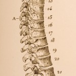
The spinal cord is protected by the spinal column, a series of bones (called vertebrae) that act as armor, protecting the spinal cord. The spinal column includes 33 bones: 7 cervical vertebrae, 12 thoracic vertebrae, 5 lumbar vertebrae, 5 sacral vertebrae, and 4 coccygeal vertebrae. The sacral and coccygeal bones will fused togethe by adulthood.
A bit of interesting developmental information: when a baby is born, the spinal cord and column is one smooth curve. Starting in the 3rd month, a baby begins to gain control of her head. This creates the first curve in the spinal cord, the cervical curve. When a baby begins to walk, the second curve, the lumbar curve, develops. Our natural spine position should include these curves as they are meant to support our upper body and head when walking upright.
Also, the spinal cord is shorter than the spinal column; it ends at the first lumbar vertebrae. The end of the spinal cord is actually a bundle of nerves that follow the column down and exit in the lumbar and sacral area. Given their appearance, this bundle of nerves is called cauda equina, or “horse’s tail.”
So, a little spinal cord anatomy for you. There is a little more spinal cord anatomy to cover (for a future post) as well as information about what to look for when damage occurs to the spine at different areas. If you know of any other information that may be pertinent or relevant for the practicing clinician, please leave a comment below!
References
Dr. Anna Fail’s Graduate Neuroanatomy class, Colorado State University
Stephen Goldberg, M.D. (2003) Clinical Neuroanatomy made ridiculously simple. Miami: MedMaster, Inc.
Dr. James Bevan (1996). A Pictoral Handbook of Anatomy and Physiology. New York: Barnes & Noble Books.


 orcid.org/0000-0001-8665-1493


{ 0 comments… add one now }
You must log in to post a comment.